Titt som tätt mejlar phi,are bloggen frågor men även kanske användbar information.Mycket av det som man vill jag ska kika närmare på är inte riktigt av den kalibern att jag tycker det är värt att uppmärksamma på bloggen.Till de som skickar sådan information återkopplar jag med anledning till icke publicerat material,det om man lämnar mejladress, Men nu har jag fått in 2 tips att kika närmare på och det har jag alltså gjort då dessa håller bra nivå.Först en forskningsrapport från Ungern,närmare bestämt från phi-kända universitetet Semmelweis.
Tyvärr hittar jag ingen engelsk version av studien så vi får hålla till godo med ungersk text.Eftersom studien är 108 sidor lång finns det ingen chans att sitta och översätta sida efter sida.Istället har jag valt ut stycken som berättar vad denna studie handlar om.Det är alltså en doktorsavhandling som då naturligtvis är tämligen gedigen och väl genomgången med referenser till 160 tidigare studier som bas.
Forskare från Semmelweis har sen tidigare specialiserat sig på åkommor som uppstår vid en graviditet.Åkommor i den bemärkelsen att graviditeten kan utlösa och resultera i allvarliga sjukdomsfall som ex cancertumörer och i värsta fall dödsfall.Söker man på Semmelweis universitet hos ställen forskare får sina studier publicerade hittar man volymer just med studier inom detta område.Exempelvis denna studie som Nature publicerade 2018. https://www.nature.com/articles/s41598-018-23706-7
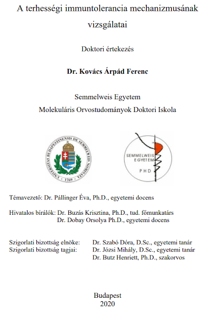
A terhességi immuntolerancia mechanizmusának vizsgálatai
The birth of a new life depends on the cooperation of two different genomes. The developing embryo and continuous lively communication between the maternal immune system is outstanding important.
One of the most dynamic forms of intercellular communication is mediated by cell membrane-surrounded blisters, i.e., extracellular vesicles (EVs) communication. EVs are evolutionarily conserved, active, unidirectional, or bidirectional create a horizontal system of connections between cells, playing a role in autocrine,in both paracrine and endocrine signaling. It is subpicomolar in the blood plasma are on average between 100 and 1 000 nm and 1 have a typical mass per gigadalt.
Their presence has been studied so far, reproductive relevant biological fluids (eg follicular fluid, amniotic fluid,breast milk, embryo culture medium, semen, blood plasma, serum). The EVs play an active role in the maturation processes, implantation and presumably in the development and maintenance of immune tolerance in relation to pregnancy is. Fetal trophoblast cells and trophoblast-derived cells direct contact of fragments / particles with maternal circulation as early as 1893 assumed.
It was later verified that it originated from the maternal-fetal interface trophoblast-derived EVs continuously enter the maternal circulation and their presence a in peripheral blood as early as the 6th week of pregnancy. From a methodological point of view,The presence of EVs can be confirmed and characterized by a flow cytometer, confocal microscopy, mass spectrometry, next generation sequencing (NGS) and by transmission electron microscopy5–9. In terms of their biochemical composition, EVs transport key regulatory proteins, lipids, and nucleic acids a cells, thereby affecting reproductive performance
Ur studien klipper jag in relevant HoloMonitorinfo.Översatt + ungerskt original.
CD3 + / CD25high + / CD127low + Treg cells were sorted for further holomicroscopy under sterile conditions (SONY SH800S) (Figure 5).
 |
| 5. ábra. In vitro differenciáltatott Treg-sejtek szortolásához alkalmazott kapuzási stratégia, FSC-H – előreszórás, SSC-H - oldalszórás |
CD25 + generated in vitro/ CD127lo Treg cells with cell sorter under sterile conditions sorted and then characterized by differentiation by holomicroscopy for 3 h cell viability. To assess viability, cell migration and motility parameters (cell pathway, motility rate) were examined. Cell migration is between 30 and 90 minutes showed the greatest heterogeneity in the study interval. Cell migration is relative showed a steady rate of cell migration (Fig. 27). By sorted Treg cells examination of the path taken, i.e., monitoring of cell migration activity is appropriate.
IEV-induced THP-1 cell migration was examined by holomicroscopy compared to untreated THP-1 cells in both preeclampsia,both healthy iEVs stimulated migration of THP-1 cells but preeclampsia
and healthy iEV effect was not significantly different (preeclampsia iEV: 0.142 ± 0.003 slope, healthy iEV: 0.139 ± 0.006 slope). However the preeclampsia iEVs induced significantly lower cell motility in the healthy compared to iEVs (preeclampsia iEV: 3.22 ± 0.008 slope, healthy iEV: 4.82 ± 0.06 slope) (Fig. 40). Pregnancy-associated EVs affect THP-1 cells.
Materials and Methods
It was placed in a Petri dish (d = 35 mm) and cultured for 24 h, then 8 x 10⁴ healthy ill. treated with iEVs isolated from the plasma of preeclampsia pregnant women. Induced by iEV migration and motility changes over 2 hours with 30-second sampling detected using the interval. Untreated THP-1 cells as a control we used. For differentiated Treg cells, 60 seconds for 3 hours measurements were performed using sampling intervals.To evaluate the images, use the automatic background threshold method (“Minimal error sets”).algorithm, cut-off = 128) was used. 50 cells were evaluated as our field of view. The 42 the number of fields of view evaluated was 240. During the assessment in the marginal zones located cells were not considered.


Inga kommentarer:
Skicka en kommentar