Neuroblastoma is a childhood solid tumor composed of primitive cells
derived from precursors of the autonomic nervous system. This neoplasm
has the highest rate of spontaneous regression of all cancer types and
has been noted to undergo spontaneous and chemically induced
differentiation into elements resembling mature nervous tissue.
Alpha-Amylase Inhibits Cell Proliferation and Glucose Uptake in Human Neuroblastoma Cell Lines
Kateryna Pierzynowska ,Sofia Thomasson,and Stina Oredsson
Published 25 Jul 2022
Abstract
The present article describes a study of the effects of alpha-amylase (α-amylase) on the human neuroblastoma (NB) cell lines SH-SY5Y, IMR-32, and LA-N-1. NB is the most common malignancy diagnosed in infants younger than 12 months. Some clinical observations revealed an inverse association between the risk of NB development and breastfeeding. α-Amylase which is present in breast milk was shown to have anticancer properties already in the beginning of the 20th century. Data presented here show that pancreatic α-amylase inhibits cell proliferation and has a direct impact on glucose uptake in the human NB cell lines. Our results point out the importance of further research which could elucidate the α-amylase mode of action and justify the presence of this enzyme in breast milk as a possible inhibitor of NB development. α-Amylase can be thus recognized as a potential safe and natural mild/host anticancer agent minimizing chemotherapy-related toxicity in the treatment of NB.
1. Introduction
Neuroblastoma (NB) is the most common extracranial paediatric tumor, and it is the most common malignancy diagnosed in infants younger than 12 months. The incidence rate of NB is almost twice that of leukaemia (the second neonatal cancer) in the mentioned paediatric group [1–4]. Regarding clinical presentation and prognosis, NB demonstrates a variety of patterns, from spontaneous regression to aggressive metastatic tumors [5, 6]. The clinical response differs dramatically in different risk groups. The same variable pattern is observed regarding the five-year survival rates, where low- and intermediate-risk cases have survival rates over 90%, while high-risk patients have a survival rate of only 40-50%, despite the intensification of treatments and incorporation of immunotherapies. It is worth mentioning that high-risk NB is found in about 40% of cases and is often accompanied with chemoresistance and tumor relapse [5, 6]. The goal of current strategies for NB treatment is to decrease therapy- and minimize chemotherapy-related toxicity for patients from low and intermediate NB risk groups, whereas concerning high risk NB cases, present and upcoming trials are devoted to the development of novel immunotherapies, inhibitors of aberrant pathways (such as MYC and ALK), and radioisotope-containing regimens.
2. Materials and Methods
2.4. Digital Holographic Imaging and Analysis
To monitor the effect of α-amylase on motility, accumulated distance, and proliferation of neuroblastoma cells, the phase holographic microscope M4 (Phase Holographic Imaging AB, Lund, Sweden) was used. Cells were detached with 0.05% trypsin/1 mM EDTA and counted in a hemocytometer. SH-SY5Y cells were seeded at a density of 20000 cells/cm2, and LA-N-1 cells were seeded at a density of 30000 cells/cm2 in 6-well plates (Nunc, Thermo Fisher Scientific). After 24 h of incubation, the cell medium was aspirated, and 4 ml of fresh cell medium was added to each well. Subsequently, three wells were treated with 2 U/l α-amylase and the three remaining wells were treated with the equivalent volumes of 0.9% NaCl. After adding α-amylase, specific HoloLids™ (Phase Holographic Imaging AB) replaced the standard 6-well plate lid and the plate was placed on the stage of the M4 HoloMonitor inside a CO2 cell culturing incubator. Images were acquired every 5 minutes for 72 h using AppSuite™ (Phase Holographic Imaging AB). Following image acquisition, the time-lapses were used to analyze kinetic motility and proliferation of single cells and videos made using AppSuite™ (Phase Holographic Imaging AB).
3.2. Cell Proliferation
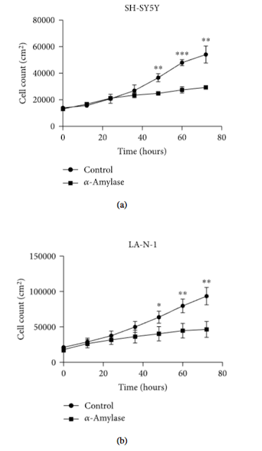
Growth curves for NB cells treated with 2 U/l α-amylase were established by digital holography. α-Amylase treatment significantly reduced number of SH-SY5Y cells by 32% () and LA-N-1 cells by 37% (), already after 48 h of incubation (Figures 2(a) and 2(b), respectively). This decline in cell count was observed until the end of the time-lapse and reached 46% () and 50% () for SH-SY5Y and LA-N-1 cells, respectively (Figure 2).
The inhibition of cell proliferation by α-amylase treatment of SH-SY5Y and LA-N-1 cells was also proven by time-lapse imaging using digital holographic microscopy (significance found after 48 h of treatment). Interestingly, the digital holographic microscopy revealed a significant effect on cell motility and accumulated distance of LA-N-1 cells, but not SH-SY5Y cells, already after 24 h of α-amylase treatment.
Som service till alla ev HoloMonitornyfikna forskare : PHIAB Webshop

Inga kommentarer:
Skicka en kommentar