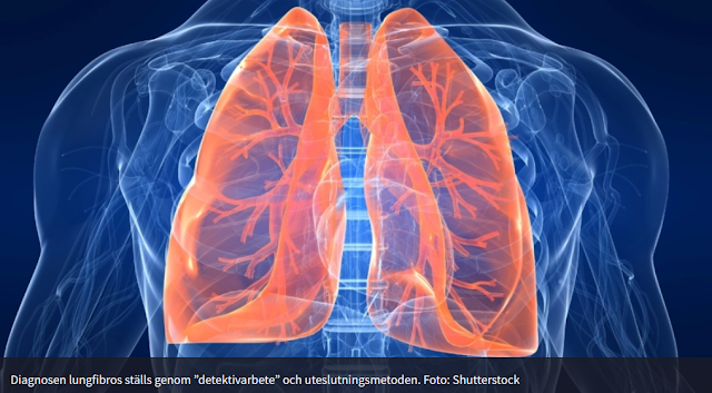
Oscar mejlar över en ny studie utförd av forskare från Lunds Universitet och Sahlgrenska Sjukhuset i Göteborg. Forskarna har studerat lungvävnad hos patienter med diagnosen idiopatisk lungfibros. En läskig sjukdom som inte sällan leder till en plågsam död. Idag finns inget botemedel mot sjukdomen,enbart en lungtransplantation kan rädda de som är svårast drabbade. Övriga behandlar man med läkemedel som förebygger och bromsar förloppet till det svåraste stadiet.
Info om sjukdomen : Allt om lungfibros (idiopatisk)
Det forskarna studerat är om en viss peptid (molekyl bestående av kroppsegna aminosyror),Dipeptidyl peptidase 4 (DPP4), kan få lungans bindvävsceller att dela sig och på det sättet läka det ärrade sjuka området.
Info :Bindvävsceller kallas även fibroblaster. Wiki berättar mer om fibroblaster.
Studiens inledning beskriver det såhär :
Dipeptidyl peptidase 4 (DPP4) has been proposed as a marker for activated fibroblasts in fibrotic disease. We aimed to investigate whether a profibrotic DPP4 phenotype is present in lung tissue from patients with idiopathic pulmonary fibrosis (IPF). The presence of DPP4+ fibroblasts in normal and IPF lung tissue was investigated using flow cytometry and immunohistology. In addition, the involvement of DPP4 in fibroblast activation was examined in vitro, using CRISPR/Cas9 mediated genetic inactivation to generate primary DPP4 knockout lung fibroblasts. We observed a reduced frequency of primary DPP4+ fibroblasts in IPF tissue using flow cytometry, and an absence of DPP4+ fibroblasts in pathohistological features of IPF. The in vivo observations were supported by results in vitro showing a decreased expression of DPP4 on normal and IPF fibroblasts after profibrotic stimuli (transforming growth factor β) and no effect on the expression of activation markers (α-smooth muscle actin, collagen I and connective tissue growth factor) upon knockout of DPP4 in lung fibroblasts with or without activation with profibrotic stimuli.
Studien som publicerades igår 31/8 är betitlad:
Dipeptidyl peptidase 4 expression is not associated with an activated fibroblast phenotype in idiopathic pulmonary fibrosis
Materials and Methods (urval)
2.8 Live cell imaging using phase holographic imaging
Cells plated at a low concentration (1300 cells/cm2) in 6-well plates (TC 6 well plate, Sarstedt) were imaged using a Holomonitor M4 live cell imaging system (Phase Holographic Imaging, PHI) placed in an incubator set at 37 °C and 5% CO2. Images from 5-8 regions per well were captured every 15 min over 72 h. Analysis of time-lapse images was performed using HStudio (PHI). To assess cell proliferation, 3-6 regions per sample (well) were used for analysis. Fold changes of cell number at different timepoints compared to 0 h for each region were calculated. The average of fold changes across each sample and timepoint were then calculated. To assess cell motility, 7–12 cells collected from 2-3 regions for each sample were tracked and analyzed over 12 h. To assess cell morphology, 5–16 cells per sample were analyzed at the 12 h time point. Elongation was calculated as the ratio between box length and box breadth of cells.
Den påläste phi,aren vet att single cell observationer är en av många HoloMonitor-funktionaliteter.
Sist några ord om våra vänner garanterna.
Mvh (en stridslysten) the99
Som service till alla ev HoloMonitornyfikna forskare : PHIAB Webshop

Inga kommentarer:
Skicka en kommentar