2 franska forskare däribland kände Monsieur Alain Géloën har lämnat in en studie för slutlig granskning innan publicering. Det handlar om....ja vad handlar studien om egentligen? Monsieur Géloën har höjt svårighetsgraden från tidigare studier så nu börjar det bli knepigt att hänga med i svängarna. Men ok..jag gör ett försök. Forskarna har undersökt hur de kroppsegna aminosyrorna glycin, cystein och glutaminsyra,förkortat GHS tillsammans, påverkar kroppens cellprocesser.
GHS är starka antioxidanter och håller kroppens celler fria från giftiga "intrång" från ex sånt som fria radikaler. Glasklart va?
Bra,då går vi vidare. GHS är även känt som en komponent för celler att dela sig och fästa i varandra (cell adhesion). Kan man få ner koncentrationen av GHS i exempelvis en cancersjuk människas kropp,påverkar det cancerns förmåga att dela sig,öka i antal cancerceller och sprida sig? Just det sökte de franska forskarna svaret på.
För att komma till svar krävdes ett speciellt instrument från lilla Sverige,närmare bestämt från Lund.
Instrumentets namn? Hehe..vad tror ni? Klart det är HoloMonitor.
Då kör vi studien och börjar med abstractet.Hela studien läses i pdf som laddas ner från sidan.
Keywords: glutathione, cell volume, cell adhesion, xCELLigence, Holomonitor, A549.
Abstract
Glutathione
is the most abundant thiol in animal cells. Reduced glutathione (GSH)
is a major intracellular antioxidant neutralizing free radicals and
detoxifying electrophiles.
It plays important roles in many cellular
processes, including cell differentiation, proliferation, and apoptosis.
In the present study we demon-strate that extracellular concentration
of reduced glutathione markedly increases cell volume within few hours,
in a dose-response manner.
Pre-incubation of cells with BSO, the
inhibitor of gamma-glutamylcysteine synthetase, responsible for the
first step in intracellular glutathione synthesis did not change the
effect of reduced glutathione on cell volume suggesting a mechanism
limited to the interaction of extracellular re-duced glutathione on cell
membrane.
Similarly, inhibition of γ-glutamylcyclotransferase involved
in intra-cellular glutamate production had no effect on the action of
reduced glutathione.
Oxidized glutathione exerted no effect on cell
volume. Results show that reduced GSH decreases cell adhesion resulting
in an increased cell volume. Since many cell types are able to export
GSH, the present results suggest that this could be a fundamental
self-regulation of cell volume, giving the cells a self-control on their
adhesion proteins.
Materials and Methods (urval)
2.3.2 Quantitative phase imaging
Quantitative phase imaging was performed using the Holomonitor M4 digital holographic
cytometer (DHC) from Phase Holographic Imaging (PHI, Lund, Sweden). The microscope was
housed in a standard 37°C cell culture incubator with 5% CO2. Average optical cell volumes were
measured in real-time every ten minutes during ten hours in control and after GSH addition.
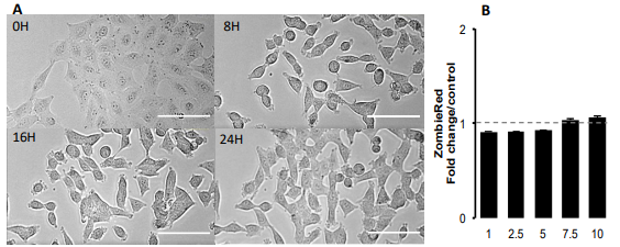
Figure 2: Morphological changes induced by glutathione in A549 cells. A: Contrast phase
photomicrographs at times 0, 8h, 16h and 24h of a time-lapse in presence of 10 mM glutathione.
No
image of dead cell is visible. Compare to time 0, cells reduced their surface. Scale bar represents
100µm. B: Cell permeability to Zombie Red analyzed by fluorescence quantification in cytometry 2.5
hours after treatment, in a dose-dependent response to glutathione (mM). Data are presented as
mean ± fold changes Zombie Red fluorescence intensity in reduced GSH treated cells versus control
media (n=4).
3.3. Effects of increasing concentrations of glutathione on cell volume.
To further quantify cellular changes observed in response to GSH, we used the label-free single-cell
analysis microscope Holomonitor (Phase Holographic Imaging, PHI, Lund, Sweden). The average
optical volume of cells significantly increased (Figure 5A). That increase was dose-dependent and
was already observable in response to 1 mM of GSH. Similar results were obtained in response to
NAC (Figure 5B)
4. Discussion.
The present results show a marked effect of GSH on cell volume. Following the addition of GSH in
the culture medium, the decrease in cell index is fast and marked reaching almost basal values,
occurring within 2-3 hours (Figure 1A). The decrease in cell index is dose-dependent to GSH
concentration (Figure 1). Similar effects are observed in response to NAC, although reduced in
amplitude compare to GSH (Figure 1B). GSH markedly reduced cell index, the visual control using
microscopy certified the absence of dead cell (Figure 2A). Furthermore, cells maintained their
membrane integrity, attested by Zombie red labeling (Figure 2B). Finally, the decrease in cell index
is reversible.
Indeed, after longer time exposure, cell indexes slowly increase despite the presence of
reduced glutathione (Figuer 3, 4 and 7). After washing out, the cell index increased markedly without
fully reaching the control values (not shown).
On xCELLigence, the cell index depends on the surface
occupied by cells but also on cell adhesion strength. Since cell number is constant the only one
explanation for the decreased cell index is that cell adhesion is affected by GSH.
Real-time analysis
using the quantitative phase contrast microscope Holomonitor M4, confirmed that GSH did not kill
cells but resulted in a marked increase in average optical cell volume (Figure 5). That increase is dosedependent. A designed experiment on xCELLigence biosensor dedicated to the measure of cell
adhesion shows a dose-response to GSH on A549 cell adhesion, thus strongly arguing for a
diminished cell adhesion force induced by GSH, which was confirmed by a reduction of F-actin
required for integrin clustering needed for cell adhesion (review in [15]).
Whether GSH regulates cell adhesion through extracellular action rather than increased intracellular
contents was supported by the extremely fast cell adhesion force reduction observed in real-time
experiments.
Indeed, a significant effect was detected within few minutes even at low concentrations
after GSH addition.
Min kommentar
HoloMonitors kompis xCELLigence från bjässen Agilent kamperar ånyo tillsammans i en ny studie signerad HoloMonitorfrälste Monsieur Géloën. (Ni vet vad jag sagt om Agilent vs PHI)
Resultatet då?
Jo,man kom fram till att minskad produktion av GHS hade påverkan på en lungcancerpatient och hanses cancer cells processer. Ännu ett framsteg i kampen mot cancer mao.
"The present study demonstrates that GSH has a major impact on cell adhesion. That decrease in cell
adhesion results in an increased cell volume. That rises the hypothesis that GSH produced by cells
can exert a direct control on cell adhesion and cell volume. Such a hypothesis adds a new function
of glutathione and most likely will necessitate a re-evaluation of cell adhesion functions according
to their redox-state."
Mvh the99


Och aktien säljs ut ännu mer...
SvaraRadera