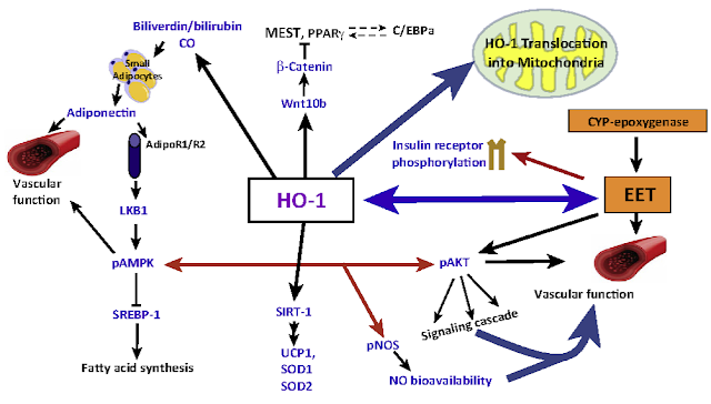
Igår publicerades en ny studie angående Systemisk Skleros.4 forskare från Leeds, England har studerat hur det kroppsegna enzymet Hemeoxygenase-1 (HO-1) påverkar en insjuknad patients hjärt och kärlsystem.
Systemisk Skleros är en dödlig sjukdom (terminal) som det idag inte finns något bra botemedel mot.
Den forskning som bedrivits i ämnet är knapp men vetenskapen har konstaterat att sjukdomen tar sin början i kroppens immunförsvar.Där sker en utveckling vilket gör att immunförsvaret ser kroppens egna vävnader som "fiender" och processer startas för att (felaktigt) påverka dessa tyvärr i en negativ riktning.
Socialstyrelsen har en bra beskrivning av sjukdomen.Från den texten klipper jag utvalda delar.
Systemisk skleros är en ovanlig bindvävssjukdom som karaktäriseras av
förändrat immunförsvar, försämrad blodcirkulation och ökad
bindvävsproduktion i hud och inre organ, framför allt i mag-tarmkanal,
lungor, hjärta och njurar. Sjukdomen kallades tidigare sklerodermi
(skleros=förhårdning, derma=hud).
Systemisk skleros är en autoimmun sjukdom, vilket innebär att kroppens
immunförsvar felaktigt riktas mot de egna vävnaderna. Trots att
grundorsaken till sjukdomen är okänd är det relativt väl känt vad som
bidrar till att den utvecklas.
Inflammatoriska celler (makrofager och lymfocyter) ansamlas kring
blodkärlen. Dessa celler utsöndrar signalsubstanser (cytokiner,
exempelvis TGFβ) som aktiverar bindvävsceller (fibroblaster) för att
dessa ska producera bindvävsmolekyler, till exempel kollagen.
Fibroblaster ska normalt aktiveras för att reparera skador, som vid
sårläkning, eller när bindvävsmolekylerna successivt ska bytas ut. Vid
systemisk skleros bildas onormalt stora mängder bindvävsproteiner i hud
och inre organ. Bindvävens normala funktion är att ge kroppens vävnader
elasticitet, stöd och hållfasthet. Organ som vid systemisk skleros
innehåller ökade mängder bindväv, som hud och lungor, blir stela och
mindre elastiska.
Onormala immunreaktioner sker, med produktion av antikroppar riktade
mot kroppsegna strukturer (autoantikroppar), till exempel mot ämnen i
cellkärnor.
Det finns ännu ingen botande behandling för systemisk skleros.
Insatserna inriktas på att lindra symtomen genom att dämpa
inflammationer och förbättra cirkulationen. Eftersom sjukdomen kan
påverka inre organ är det viktigt att organfunktionen undersöks noga och
att vård och behandling ges tidigt i
sjukdomsförloppet.
Men till de engelska forskarnas studie.
Front. Physiol., 05 May 2022
Systemic sclerosis (SSc) is a terminal disease characterized by
vasculopathy, tissue fibrosis, and autoimmunity. Although the exact
etiology of SSc remains unknown, endothelial dysfunction, oxidative
stress, and calcium handling dysregulation have been associated with a
large number of SSc-related complications such as neointima formation,
vasculogenesis, pulmonary arterial hypertension, impaired angiogenesis,
and cardiac arrhythmias. Hemeoxygenase-1 (HO-1) is an antioxidant enzyme
involved in multiple biological actions in the cardiovascular system
including vascular tone, angiogenesis, cellular proliferation,
apoptosis, and oxidative stress. The aim of this work was to investigate
the physiological role of HO-1 and its relevance in the cardiovascular
complications occurring in SSc. We found that, in early phases of SSc,
the expression of HO-1 in dermal fibroblast is lower compared to those
isolated from healthy control individuals. This is particularly relevant
as reduction of the HO-1/CO signaling pathway is associated with
endothelial dysfunction and vasculopathy. We show evidence of the role
of HO-1/carbon monoxide (CO) signaling pathway in calcium handling.
Using an in vitro model of pulmonary arterial hypertension (PAH) we investigated the role of HO-1 in Ca2+
mobilization from intracellular stores. Our results indicate that HO-1
regulates calcium release from intracellular stores of human pulmonary
arterial endothelial cells. We interrogated the activity of HO-1 in
angiogenesis using an organotypic co-culture of fibroblast-endothelial
cell. Inhibition of HO-1 significantly reduced the ability of
endothelial cells to form tubules. We further investigated if this could
be associated with cell motility or migration of endothelial cells into
the extracellular matrix synthesized by fibroblasts.
By mean of
holographic imaging, we studied the morphological and functional
features of endothelial cells in the presence of an HO-1 activator and
selective inhibitors. Our results demonstrate that inhibition of HO-1
significantly reduces cell proliferation and cell motility (migration)
of cultured endothelial cells, whilst activation of HO-1 does not modify
either morphology, proliferation or motility. In addition, we
investigated the actions of CO on the Kv7.1 (KCQN1) channel current, an
important component of the cardiac action potential repolarization.
Using electrophysiology (whole-cell patch-clamp in a recombinant system
overexpressing the KCQN1 channel), we assessed the regulation of KCQN1
by CO. CORM-2, a CO donor, significantly reduced the Kv7.1 current,
suggesting that HO-1/CO signaling may play a role in the modulation of
the cardiac action potential via regulation of this ion channel. In
summary, our results indicate a clear link between: 1) downregulation of
HO-1/CO signaling; and 2) pathophysiological processes occurring in
early phases of SSc, such as calcium homeostasis dysregulation, impaired
angiogenesis and cardiac arrhythmias. A better understanding of the
canonical actions (mainly due to the biological actions of CO), and
non-canonical actions of HO-1, as well as the interaction of HO-1/CO
signaling with other gasotransmitters in SSc will contribute to the
development of novel therapeutic approaches.
Materials and Methods (urval)
Live Holographic Imaging
Holographic
microscopy analysis was performed to investigate the morphological and
functional changes caused by inducers and inhibitors of HO-1 in
endothelial cells. Holographic imaging recorded in real-time was used to
monitor morphological (cell area, optical thickness, and optical
volume) and functional (cell motility, cell tracking, and cell
migration) parameters using the Holomonitor App Suite (Phase Holographic
Imaging PHI AB). HUVECs were seeded at low confluency (100 k cells per
well) onto 6-well plates (Sarstedt, Germany). Drugs and VEGF were added
to the media at the concentration indicated in Figure 4
(25 ng/ml VEGF, 10 µM CoPPIX, and 1 µM ZnPPIX). For every independent
experiment, one control well (absence of drugs or VEGF) was included in
each plate.
The plate was placed onto the xy motorised stage of a
HoloMonitor M4 Microscope, set up inside an incubator using Phi
HoloLids™ imaging covers (PHI, phase holographic imaging), previously
sterilised in 70% EtOH, to optimise the acquisition quality of the
holographic images. After automatic calibration of the background and
microscope objective, a minimum of three fields/coordinates per well,
randomly assigned by the software, were focused on. Holographic images
were recorded at an interval of 5 min over 48 h using the HoloMonitor M4
(Phase Holographic Imaging PHI AB, Lund, Sweden) inside the
cell-culture incubator (37°C, 5% CO2). Post-acquisition, the
images were analysed applying the “Otsu mask”, where a threshold was set
to distinguish cells from the bottom of the well (cell segmentation),
allowing for automatic cell identification. Automatic cell number
assignment allowed for individual cell tracking over time, which was
double-checked manually as described in (Owston et al., 2019). All outputs were exported into Excel files and plotted using Prism 9 (GraphPad).
Resultatet forskarna kom fram till beskrivs enligt följande :
- As fibroblasts are responsible for the production and secretion of TGF-β
(the main pro-fibrotic factor in SSc), we investigated the expression
levels of HO-1 in healthy and SSc fibroblasts. Although expression
levels of HO-1 in unstimulated SSc fibroblasts are not significantly
different from healthy fibroblasts, exposure to 10 µM TGF-β for 24 h
significantly inhibited the expression of HO-1 in healthy but not SSc
fibroblasts (Figure 1A),
indicating that the expression of HO-1 is already influenced by the
elevated endogenous production of TGF-β in SSc fibroblasts.
Glasklart, eller hur?
Forskarna avslutar rapporten med att tacka PHI`s repr Amendeep Dhillon.
Acknowledgments
We are extremely grateful to Amandeep Dhillon (Phase Holographic Imaging
PHI AB) for lending the University of Bradford the HoloMonitor® M4 to conduct the cellular morphology and migration studies.
Min kommentar
Ånyo ett nytt användningsområde för HoloMonitor kan denna studie visa på.Tekniken firar segrar medan (aktie)marknaden fortfarande surar.Dock finns små tecken på att det kommer förändras inom kort.
Mvh the99
Som service till alla ev HoloMonitornyfikna forskare : PHIAB Webshop

Inga kommentarer:
Skicka en kommentar